SCHEDULE YOUR APPOINTMENT
CALL: 224-723-5588
SAME DAY APPOINTMENTS AVAILABLE
RECONSTRUCTIVE REARFOOT
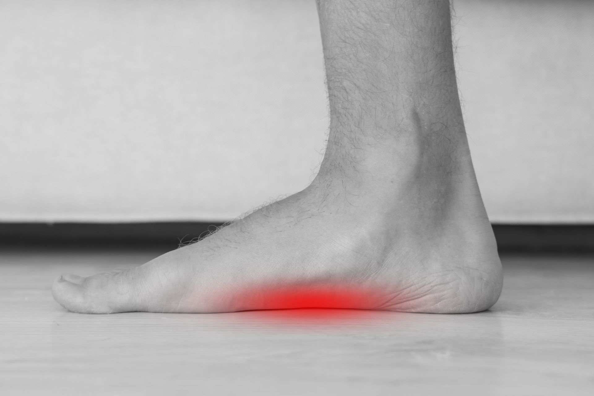
Flatfoot and cavus foot are deformities that affect the arch of the foot, with flatfoot causing a collapsed arch and cavus foot creating a high arch, both leading to pain, instability, and potential complications.
Both conditions may require treatment ranging from orthotics and bracing to surgery, depending on the severity and underlying causes.
Flatfoot
This condition is characterized by the inability to lift the front part of the foot, causing scuffing or dragging while walking. It is typically caused by nerve or muscle disorders. Dr. Levick offers treatments to improve gait and mobility.
High Arched Feet
Common in young children, toe walking can be idiopathic or related to a shortened Achilles tendon. If not outgrown in early childhood, an evaluation is necessary to prevent future complications. Dr. Levick provides expert care for addressing toe walking concerns.
What is Flatfoot?
Flatfoot is often a complex disorder, with diverse symptoms and varying degrees of deformity and disability. There are several types of flatfoot, all of which have one characteristic in common: partial or total collapse (loss) of the arch. Other characteristics shared by most types of flatfoot include toe drift, in which the toes and front part of the foot point outward. The heel tilts toward the outside and the ankle appears to turn in. A tight Achilles tendon, which causes the heel to lift off the ground earlier when walking and may make the problem worse, bunions and hammertoes may develop as a result of a flatfoot.
In some cases, a tarsal coalition, or a connection between two bones (by bone, cartilage or fibrous tissue) that shouldn’t be there can be the culprit of a pediatric flatfoot. Dr. Levick will be able to resect the coalition depending on the % of the joint affected.
Symptoms
Symptoms that may occur in some persons with flexible flatfoot include:
- Pain in the heel, arch, ankle or along the outside of the foot
- Rolled-in ankle (overpronation)
- Pain along the shin bone (shin splint)
- General aching or fatigue in the foot or leg
- Low back, hip or knee pain
Although most patients respond to conservative treatment, surgery may be considered after failing custom orthotics, bracing and physical therapy.
Many surgical techniques are available to correct flexible flatfoot, and one or a combination of procedures may be required to relieve the symptoms and improve foot function. In selecting the procedure or combination of procedures for your particular case, Dr. Levick will take into consideration the extent of your deformity based on the x-ray findings, your age, your activity level and other factors. The length of the recovery period will vary depending on the procedure or procedures performed.
What Is Cavus Foot?
Cavus foot is a condition in which the foot has a very high arch. The high-arched foot places an excessive amount of weight on the ball and heel of the foot when walking or standing. Cavus foot can lead to a variety of signs and symptoms, such as pain and instability. It can develop at any age and can occur in one or both feet.
Cavus foot is often caused by a neurologic disorder or other medical condition, such as cerebral palsy, Charcot-Marie-Tooth disease, spina bifida, polio, muscular dystrophy or stroke. In other cases of cavus foot, the high arch may represent an inherited structural abnormality. An accurate diagnosis is important because the underlying cause of cavus foot largely determines its future course. If the high arch is due to a neurologic disorder or other medical condition, it is likely to progressively worsen. On the other hand, cases of cavus foot that do not result from neurologic disorders usually do not change in appearance.
Symptoms of Cavus Foot (High-Arched Foot)
The arch of a cavus foot will appear high even when standing. In addition, one or more of the following symptoms may be present:
- Hammertoes or claw toes
- Calluses on the ball, side or heel of the foot
- Outside of foot pain
- Chronic ankle sprains
- Bracing. The surgeon may recommend a brace to help keep the foot and ankle stable. Bracing is also useful in managing foot drop.
If nonsurgical treatment fails to adequately relieve pain and improve stability, surgery may be needed to decrease pain, increase stability and compensate for weakness in the foot.
Dr. Levick will choose the best surgical procedure or combination of procedures based on the patient’s individual case. In some cases where an underlying neurologic problem exists, surgery may be needed again in the future due to the progression of the disorder.

1535 Lake Cook Road Suite 206
Northbrook IL 60062
HOSPITAL AFFILIATIONS
- Advocate Good Shepherd Hospital
- Endeavor Health System (Skokie, Evanston, Glenbrook and Highland Park Hospitals)
- Northwest Community Hospital
CONTACT
Created by Mindset Media Group. The content on this website is owned by us and our licensors. Do not copy any content (including images) without our consent.


